Case Report
2015
March
Volume : 3
Issue : 1
Ectodermal dysplasia and partial anodontia: A case report
Prasad M, Prathyusha M
Pdf Page Numbers :- 40-42
Prasad M1,* and Prathyusha1
1Great Eastern Medical School and Hospital, Ragolu, Srikakulam-532484, Andhra Pradesh
2Dental Care, Krishna Institute of Medical Sciences, Minister Road, Secunderabad-500003, Telangana
*Corresponding author: Dr. M. Prasad, MDS, Great Eastern Medical School and Hospital, Ragolu, Srikakulam-532484, Andhra Pradesh.
Received 11 November 2014; Revised 10 December 2014; Accepted 17 December 2014; Published 21 December 2014;
Citation: Prasad M, Prathyusha. Ectodermal dysplasia and partial anodontia: A case report. J Med Sci Res 2015; 3(1):40-42. DOI: http://dx.doi.org/10.17727/JMSR.2015/3-008
Copyright: © 2015 Prasad M, et al. Published by KIMS Foundation and Research Center. This is an open-access article distributed under the terms of the Creative Commons Attribution License, which permits unrestricted use, distribution, and reproduction in any medium, provided the original author and source are credited.
Abstract
Ectodermal dysplasia is a hereditary disorder that occurs as a consequence of disturbances in the ectoderm of the developing embryo. It is usually accompanied by lack of sweat glands and a partial or complete absence of primary and/or permanent dentition. A case report illustrating the prosthetic rehabilitation of a young woman with anhidrotic ectodermal dysplasia associated with partial anodontia and mid facial defect are presented. Since the oral rehabilitation of these cases is often difficult; treatment should be administered by a multidisciplinary team involving orthodontics, prosthodontics and oral-maxillofacial surgery.
Keywords: Ectodermal dysplasia; hereditary disorder; anodontia
Full Text
Ectodermal dysplasia syndrome (EDS) is a large, heterogeneous group of inherited disorders, the manifestations of which could be seen in more than one derivative. These tissues primarily are the skin, hair, eccrine glands, and teeth [1-3]. Ectodermal dysplasia represents a large and complex group of diseases comprising more than 170 different clinical conditions. The incidence of this condition is 1:100,000 when at least 2 types of abnormal ectodermal features occur, such as malformed teeth and extremely sparse hair, the patient is diagnosed with ectodermal dysplasia syndrome [4-8].
The current classification of EDS is based on clinical features. Most commonly EDS is of two types i.e. Hypohidrotic (anhidrotic) ED (christ-siemen-touraine syndrome) and Hidrotic ED (clouston syndrome).
Clinical features
Generally includes reduction of hair follicles varying from sparse scalp hair to complete absence of hair. Eccrine glands may be absent or rudimentary. Mouth may be dry from hypoplasia of salivary glands, lacrimal glands may be deficient. Nails are often brittle and thin. Oral traits of ectodermal dysplasia (ED) may be expressed as anodontia or hypodontia, with or without a cleft lip and palate. Anodontia also manifests itself by a lack of alveolar ridge development; 7 as a result, the vertical dimension of the lower face is reduced, the vermilion border disappears, existing teeth are malformed, the oral mucosa becomes dry, and the lips become prominent. The face of an affected child usually has the appearance of old age.
Case presentation
A 22-year-old female patient reported to the department of dental care, KIMS hospital, Secunderabad, with the complaint of multiple missing teeth since childhood (Figure 1). She underwent surgery for the cleft palate during childhood. The patient also gave a history of delay in the eruption of deciduous and permanent teeth, intolerance to heat and reportedly less sweat production. There was no history of consanguineous marriage between the parents. On extra oral examination, the patient had dry skin with periocular area being hyper pigmented and wrinkled with sparse hair on the body and scalp. Hairs present were fine in texture & lighter in color. Prominent supraorbital ridges, frontal bossing, small and outwardly placed ears and flattened nasal bridge was also present. Both upper and lower eyelids showed sparse eyelashes. The skin was warm and dry, and absence of tears was also reported.

|
Figure 1: Front & Lateral view of patient with ectodermal dysplasia.
|
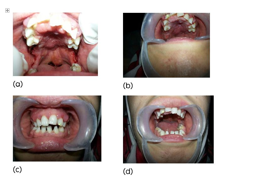
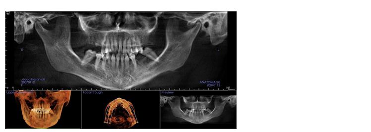
Intra oral examination revealed multiple missing teeth (Figure 2a,b,c,d) in the maxillary and mandibular arches, malalignment of teeth was present, palatal fistula with narrow maxillary arch seen, and in addition to that swollen gums were present. Based on these findings a diagnosis of ectodermal dysplasia was made. A cone-beam computed tomography (CBCT) scan was made which revealed multiple missing teeth (Figure 3), inadequate bone density in certain areas.

Figure 2a-d: Intra-oral view of patient showing partial anodontia and conical teeth.

|
Figure 3: Pan view of CBCT scan and showing multiple missing teeth.
|
Accordingly her treatment was planned in three phases, which are i) Laser gingivoplasty for inflamed and swollen gums, ii) Orthodontic correction of malaligned teeth followed by closure of the palatal fistula with the intervention of plastic surgeon, iii) Replacement of missing teeth with implants and crowns.
After taking the consent from the patient and her family, laser treatment was done for the gums, orthodontic treatment was done with periodic evaluation to align the teeth. Along with the orthodontic correction 2 stage implants were placed in areas of missing teeth. A period of 6 months was required for proper osseo integration of the implants and proper healing of gums.
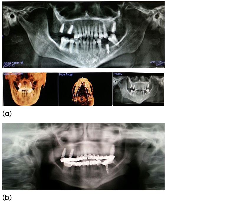
After 6 months, a CBCT scan was taken again and proper osseo integration of implants was seen, teeth were aligned and the fistula was closed. Therefore crowns were placed to replace the missing teeth (Figure 4). The facial profile and expression improved significantly (Figure 5).

|
Figure 4a, b: Post operative orthopantamogram
|

Conclusion
Ectodermal dysplasia is a rare genetic disorder with involvement of various tissues in the body. A careful and a thorough examination will lead to accurate diagnosis. Restoration of normal function should be the main concern in these patients.
Acknowledgements
Acknowledgements are due to the Krishna Institute of Medical Sciences (KIMS), Minister Road, Secunderabad - 500003, Telangana.
Conflict of interest
The authors declare no conflict of interest.
References
[1] Monreal AW, Ferguson BM, Headon DJ, Street SL, Overbeek PA, et al. Mutations in the human homologue of mouse dl cause autosomal recessive and dominant hypohidrotic ectodermal dysplasia. Nat Genet. 1999; 22(4):366–369.
[2] Kere J, Srivastava AK, Montonen O, Zonana J, Thomas N, et al. X-linked anhidrotic (hypohidrotic) ectodermal dysplasia is caused by mutation in a novel transmembrane protein. Nat Genet. 1996; 13(4):409–416.
[3] Monreal AW, Zonana J, Ferguson B. Identification of a new splice form of the EDA1 gene permits detection of nearly all X-linked hypohidrotic ectodermal dysplasia mutations. Am J Hum Genet. 1998; 63(2):380–389.
[4] Lamartine J, Munhoz Essenfelder G, Kibar Z, Lanneluc I, Callouet E, et al. Mutations in GJB6 cause hidrotic ectodermal dysplasia. Nat Genet. 2000; 26(2):142–144.
[5] Smith FJ, Jonkman MF, van Goor H, Coleman CM, Covello SP, et al. A mutation in human keratin K6b produces a phenocopy of the K17 disorder pachyonychia congenita type 2. Hum Mol Genet. 1998; 7(7):1143–1148.
[6] McLean WHI, Rugg EL, Lunny DP, Morley SM, Lane EB, et al. Keratin 16 and keratin 17 mutations cause pachyonychia congenital. Nature Genetics 1995; 9, 273–278.
[7] Bowden PE, Haley JL, Kansky A, Rothnagel JA, Jones DO, et al. Mutation of a type II keratin gene (K6a) in pachyonychia congenita. Nat Genet. 1995; 10(3):363–365.
[8] Pinheiro M, Freire-Maia N. Ectodermal dysplasias: a clinical classification and a causal review. Am J Med Genet. 1994; 53(2):153–162.